Myocardial Infarction: if the rapid development of myocardial necrosis by a critical imbalance

Pathophysiology
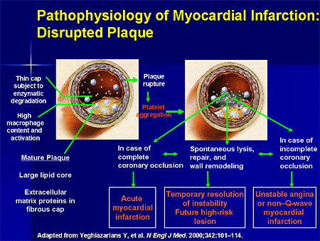
Myocardial infraction commonly known as heart attack, is the interruption of blood supply to a part of heart causing heart cells to die. This is the most commonly due to occulusion(blockage) of coronary arery following the rupture of vulnerable atherosclerotic plaque which is unstable collection of lipids(fatty acid) and white blood cell especially macrophages in the wall of an artery . The resulting ischemia ( restriction in blood supply ) and oxygen shortage. It left untreated for a sufficent period of time can cause damage or death of heart muscle tissue.
Athherosclerosis is the disease primarily responsible for most acute coronary syndrome. Approximately 90% of MI result from an acute thrombus that obstructs an atherosclerotic coronary artery. Plaque rupture is considered to be major trigger of coronary thrombosis. Following plaque rupture , platelet activation and aggregation pathway activation and endothelial vasoconstriction occurs and lead to coronary thrombosis and occlusion and eventually leads to myocardial infraction occurs.
Atherosclerotic
plaques
Plaque rupture /
erosion
↓
Thrombus formation
Embolisation
↓
Coronary vessels
obstruction
Myocardial Infraction
Classification
1. Type 1 – spontaneous MI related to ischemia due to a primary coronary event such as plaque erosion and/or rupture, fissuring, or dissection
2. Type 2 – MI secondary to ischemia due to either increased oxygen demand or decreased supply, e.g. coronary artery spasm, coronary embolism, anemia, arrhythmias, hypertension, or hypotenion
3. Type 3 – sudden unexpected cardiac death, including cardiac arrest, often with symptoms suggestive of myocardial ischemia, accompanied by new ST elevation, or new left bundle branch block (LBBB), or evidence of fresh thrombus in a coronary artery by angiography and/or at autopsy, but death occurring before blood samples could be obtained, or at a time before the appearance of cardiac biomarkers in the blood
4. Type 4 – associated with coronary angioplasty or stents:
a. Type 4a – MI associated with percutaneous coronary intervention (PCI)
b. Type 4b – MI associated with stent thrombosis as documented by angiography or at autopsy
5. Type 5 – MI associated with CABG
Also classified in two basic types of MI
a. Transmural:- infraction extends through the whole thickness of the heart muscle and are usually a result of complete occlusion of area’s blood supply
b. Subendocardial:- : involving a small area in the subendocardial wall of the left ventricle, ventricular septum, or papillary muscles. Subendocardial infarcts are thought to be a result of locally decreased blood supply, possibly from a narrowing of the coronary arteries.
Etiology
I. Hypertension
II. Unhealthy cholesterol level
III. Tobacco smoking
IV. Diabetes
V. Overweight or obese
VI. Metabolic syndrome sedentary lifestyle
VII. Age >45 for men and >55 for women.
 Clinical Features
MI patient may present with following symptoms
Prolonged cardiac pain
Chest, throat, arms, epigastrium or back
Anxiety and fear of impending death
Nausea and vomiting
Breathlessness
Collapse/syncope
Signs
1. Signs of sympathetic activation
a. Pallor, sweating, tachycardia
2. Signs of vagal activation
a. Vomiting, bradycardia
3. Signs of impaired myocardial function
a. Hypotension, oliguria, cold peripheries
b. Narrow pulse pressure
c. Raised jugular venous pressure
d. Third heart sound
e. Quiet first heart sound
f. Diffuse apical impulse
g. Lung crepitations
4. Signs of tissue damage
a. Fever
5. Signs of complications, e.g. mitral regurgitation, pericarditis
Diagnosis
✎Histry and physical examination
✎Electrocardiogram:
 High probability include ST segment elevation in two contiguous leads or presence of Q waves Intermediate probability ST depression
✎Echocardiogram
i. Use 2-dimentional and M mode echocardiography when evaluating overall ventricular function and wall motion abnormalities
ii. Echocardiography can also identify complications of MI
✎Chest X-ray
✎Routine blood test
✎ Cardiac Enzymes: creatinine kinase CK-MP, Troponin (Diagnostic), LDH & CRP.
✎Coronary angiography
✎Cardiac biomarker
a) Cardiac biomarkers are protein molecules released into the blood stream from damaged heart muscle
b) Since ECG can be inconclusive , biomarkers are frequently used to evaluate for myocardial injury
c) These biomarkers have a characteristic rise and fall pattern
Treatment
The goals of therapy in AMI are the expedient restoration of normal coronary flow and the maximum salvage of functional myocardium
1. Pharmacological Intervention
✔Pain control drugs to reduce catecholamine-induced oxygen demand to injured heart muscle.
✔Opiate analgesics: Morphine
✔Vasodilators: Nitroglycerin
✔Anxiolytics: Benzodiazepines
2. Thrombolytic therapy by I.V. or intracoronary route, to dissolve thrombus formation and reduce the size of the infarction.
✔Anticoagulants or other anti-platelet medications such as adjunct to thrombolytic therapy.
✔Reperfusion arrhythmias may follow successful therapy.
✔Beta-adrenergic blockers, to improve oxygen supply and demand, decrease sympathetic stimulation to the heart, promote blood flow in the small vessels of the heart, and provide antiarrhythmic effects.
✔Calcium channel blockers, to improve oxygen supply and demand.

Complication
Acute complications
a) Heart failure
b) Aneurysm of the left ventricle myocardium;
c) Ventricular septal rupture or free wall rupture
d) Mitral regurgitation
e) Dressler's syndrome
f) Ventricular fibrillation
g) Ventricular tachycardia
h) Atrial fibrillation, and heart block
Longer-term complications
a) Heart failure
b) Atrial fibrillation
c) Increased risk of a second MI
References
1. Wikipedia
2. Davidson's Principles and Practice of Medicine 20th & 21st Ed